The transmission electron microscope (TEM) is a scientific instrument that uses electrons instead of light to scrutinize objects at very fine resolutions. They were developed in the 1930s when scientists realized that electrons can be used instead of light to "magnify" objects or specimens under study.
TEMs provided a means to go beyond the magnification and resolution limits of light microscopes, allowing for magnification of up to 100,000x and resolutions in the nanometer range.
Uses of the Transmission Electron Microscope
The TEM has its primary uses in metallurgy (or the study of metals and minerals) and the biological sciences, especially in the study of cells at the molecular level. TEMs have been particularly useful in metallurgy, especially in terms of developing images of crystals and metals at the molecular level – allowing scientists to study their structure, interactions and identify flaws.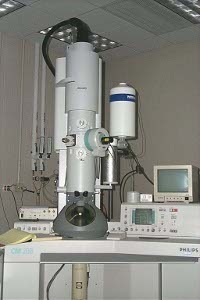
The downside to the TEM lies in the specimens that can be studied – these have to be 'sliced' very, very thinly to ensure that they are 'electron transparent'; they must also be placed in a vacuum. As such, preparation of specimens is often very time consuming and requires expert handling. This leads to concerns that specimens prepared for TEM study will be inadvertently damaged in the process. This raises the question of whether the specimen is as pure as can be expected. Finally, there are also concerns that the bombardment of electrons may damage the specimen under scrutiny – especially if these are biological samples.
How a Transmssion Electron Microscope Works
An old-fashioned slide projector works by projecting light through a film layer. As the light passes through the film, it interacts with the film and specific areas of the film lets light pass through unobstructed, other areas absorb light and doesn't let it pass through, and still some absorb part of the light and lets only a fraction get through. The light that does go through is hits the lenses found on the other side and the resulting image is projected onto a screen.
The TEM works similarly. In the case of the TEM, though, a beam of electrons are focused on a single, pinpoint spot or element on the sample being studied. The electrons interact with the sample and only those that go past unobstructed hit the phosphor screen on the other side. At this point, the electrons are converted to light and an image is formed.
The dark areas of the image correspond to areas on the specimen where fewer electrons were able to pass through (either absorbed or scattered upon impact); the lighter areas are where more electrons did pass through, although the varying amounts of electrons in these areas enable the user to see structures and gradients.
The 'lenses' in a TEM are not the same as lenses in a conventional microscope; these are actually EM devices that can 'focus' the electron beam to the desired wavelength or size. In much the same way as a light microscope, however, the amount of power used to generate electrons allows for higher magnification or better resolutions.

Follow Us!